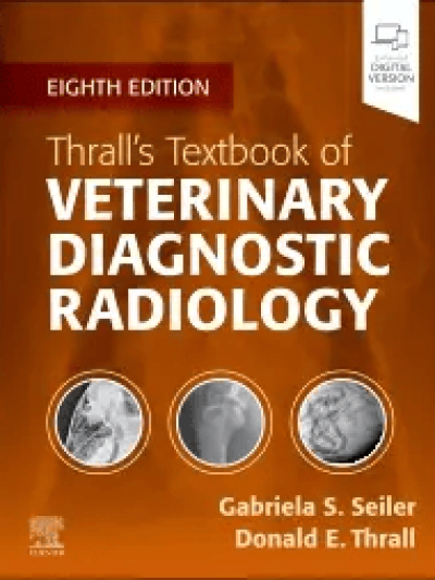
Libro: Thrall’s Textbook of Veterinary Diagnostic Radiology 8th Edition
- UPDATED! User-friendly content helps you develop essential skills in patient positioning, radiographic technique and safety measures, normal and abnormal anatomy, radiographic viewing and interpretation, and alternative imaging modalities.
- NEW! The latest digital imaging information helps you stay up to date with the latest advances in the field.
- NEW! An ebook version, included with every new print purchase, provides access to all the text, figures, and references, with the ability to search, customize content, make notes and highlights, and have content read aloud. Also included are videos, quizzes, and additional image examples of the most common diseases.
- UPDATED! Current coverage of the principles of radiographic technique and interpretation for the most seen species in private veterinary practices and veterinary teaching hospitals includes the cat, dog, and horse.
- Coverage of special imaging procedures such as the esophagram, upper GI examination, excretory urography, and cystography, helps in determining when and how these procedures are performed in today’s practice.
- Content on abdominal ultrasound imaging helps in deciding on a diagnostic plan and interpreting common ultrasound findings.
- An atlas of normal radiographic anatomy in each section makes it easier to recognize abnormal radiographic findings.
- High-quality radiographic images clarify key concepts and interpretation principles.
-
Cover image
-
Title page
-
Table of Contents
-
Copyright
-
List of Contributors
-
Preface
-
Acknowledgement
-
Video Table of Contents
-
Section I. Physics and Principles of Interpretation
-
Chapter 1. Radiation Protection and Physics of Diagnostic Radiology
-
Basic Properties of X-Rays
-
Radiation Protection
-
Production Of X-Rays
-
Interaction Of Radiation With Matter
-
Basic Concept Of Making A Radiograph
-
Factors Affecting Image Detail
-
Factors Affecting Contrast
-
Chapter 2. Digital Radiographic Imaging
-
What Is Digital Radiographic Imaging?
-
The Digital Image File
-
The Components Of A Digital Image
-
Digital Radiography Acquisition Hardware
-
Image Processing And Viewing
-
Digital Versus Analog Imaging
-
Artifacts
-
Chapter 3. Physics of Ultrasound Imaging
-
Physical Principles Of Ultrasound Waves
-
Ultrasound Wave Interaction With Matter
-
Transducers
-
Display
-
Basic Scanner Controls
-
Principles Of Interpretation
-
Doppler Techniques
-
Doppler Modes
-
Artifacts
-
Doppler Artifacts
-
Chapter 4. Principles of Computed Tomography and Magnetic Resonance Imaging
-
The Role of Computed Tomography and Magnetic Resonance Imaging in Veterinary Practice
-
Image Formation: General Concepts
-
Computed Tomography
-
Cone-Beam Computed Tomography
-
Magnetic Resonance Imaging
-
Chapter 5. Contrast Media in Diagnostic Imaging
-
Introduction
-
Radiographic Contrast Media
-
Barium Contrast Media
-
Iodine-Based Contrast Media
-
Negative Contrast Media
-
Magnetic Resonance Imaging Contrast Media
-
Ultrasound Contrast Media
-
Acknowledgment
-
Chapter 6. Radiographic Interpretation
-
What Is A Radiograph?
-
How X-Ray Exposure Is Represented in The Image
-
Radiographic Interpretation
-
Naming and Viewing Radiographs
-
Radiographic Opacities
-
Radiographic Contrast
-
Describing Abnormalities
-
Radiographic Geometry
-
Chapter 7. Errors and Pitfalls in Radiographic Interpretation
-
How We See
-
How We Think
-
Errors
-
Error Reduction
-
Section II. The Axial Skeleton: Canine, Feline, and Equine
-
Chapter 8. Radiographic Anatomy of the Axial Skeleton
-
Chapter 9. Basic Principles of Radiographic Interpretation of the Axial Skeleton
-
Skull
-
Spine
-
Chapter 10. Canine and Feline Dental Disease
-
Anatomy And Radiographic Interpretation
-
Periodontal Disease
-
Endodontic Disease
-
Tooth Resorption
-
Dentoalveolar Trauma
-
Neoplastic Disorders
-
Developmental Abnormalities Of The Teeth
-
Acknowledgment
-
Chapter 11. The Skull and Nasal Cavities: Canine and Feline
-
Normal Anatomy
-
Congenital Anomalies
-
Metabolic Anomalies
-
Neoplastic Abnormalities
-
Inflammatory and Infectious Disorders
-
Traumatic Injuries
-
Miscellaneous Diseases
-
Chapter 12. Magnetic Resonance Imaging and Computed Tomographic Features of Brain Disease in Small Animals
-
Basic Magnetic Resonance Examination Of The Brain
-
Basic Computed Tomographic Examination Of The Brain
-
Miscellaneous Disorders
-
Brain Neoplasia
-
Vascular Disruptions
-
Chapter 13. The Equine Head
-
Radiology Versus Other Imaging Modalities
-
Diseases Of The Nasal Cavity/Paranasal Sinuses
-
Dental Disease
-
Nondental Diseases Of The Mandible And Maxilla
-
Guttural Pouch
-
Larynx/Upper Airway
-
Ear/Hyoid Apparatus
-
Brain
-
Other Conditions Of The Head
-
Acknowledgment
-
Chapter 14. Radiography and Myelography of the Canine and Feline Vertebrae
-
Anatomic Considerations
-
Anomalies Of The Vertebral Column
-
Fracture And Luxation
-
Intervertebral Disc Disease
-
Inflammatory Conditions
-
Degenerative Conditions
-
Vertebral Neoplasia
-
Metabolic And Unclassified Conditions
-
Myelography
-
Acknowledgment
-
Chapter 15. Magnetic Resonance Imaging and Computed Tomography Features of Canine and Feline Spinal Cord Disease
-
Normal Appearance Of The Spine On Computed Tomography And Magnetic Resonance Imaging
-
Intervertebral Disc Disease
-
Cervical Spondylomyelopathy
-
Cystic Changes Of The Spine
-
Spinal Tumors
-
Myelomalacia
-
Vascular Pathology
-
Spinal Trauma
-
Inflammatory/Infectious Conditions
-
Vertebral Anomalies
-
Syringomyelia
-
Section III. The Appendicular Skeleton: Canine, Feline, and Equine
-
Chapter 16. Radiographic Anatomy of the Appendicular Skeleton
-
Chapter 17. Principles of Radiographic Interpretation of the Appendicular Skeleton and Radiographic Features of Bone Tumors
-
Positioning: Dog and Cat
-
Positioning: Horse
-
Aggressive Versus Nonaggressive Bone Lesions
-
Chapter 18. Orthopedic Diseases of Young and Growing Dogs and Cats
-
Disorders Primarily Affecting Joints
-
Epiphyseal Dysplasia
-
Diseases Primarily Affecting The Physis
-
Diseases Of The Metaphysis
-
Disease Primarily Affecting The Diaphysis
-
Generalized Bone Disorders
-
Focal Agenesis or Malformations
-
Diseases Primarily Affecting The Skull
-
Acknowledgment
-
Chapter 19. Fracture Healing and Complications
-
Physiology of Bone Development and Healing
-
Fracture Identification And Description
-
Fracture Healing
-
Radiographic Evaluation Of Fracture Healing And Associated Complications
-
Chapter 20. Radiographic Signs of Joint Disease in Dogs and Cats
-
Radiographic Signs Of Joint Disease
-
Sesamoid Bones
-
Degenerative Joint Disease
-
Trauma To The Joint
-
The Synovium
-
Immune-Mediated Polyarthropathies
-
Septic Arthritis
-
Miscellaneous Polyarthropathies
-
Joint Neoplasia
-
Chapter 21. The Equine Stifle
-
The Stifle
-
Diseases Of The Femoropatellar Joint
-
Diseases Of The Femorotibial Joints
-
Fractures Involving The Stifle
-
Miscellaneous Conditions Involving The Stifle
-
Chapter 22. Equine Tarsus
-
Tarsal Anatomy
-
Tarsal Radiography
-
Developmental Osseous Conditions
-
Degenerative And Acquired Osseous Conditions
-
Soft Tissue Conditions Of The Equine Tarsus
-
Acknowledgment
-
Chapter 23. Equine Carpus
-
Radiographic Study Acquisition
-
Normal Anatomical Variants And Incidental Findings
-
Radiographic Abnormalities
-
Acknowledgment
-
Chapter 24. Equine Metacarpus and Metatarsus
-
Radiographic Anatomy And Common Variants
-
Common Radiographic Abnormalities Of The Metacarpus
-
Other Imaging Techniques
-
Acknowledgment
-
Chapter 25. Equine Fetlock Joint
-
Anatomy
-
Radiographic Examination
-
Alternative Imaging Modalities
-
Radiographic Interpretation Of Diseases Of The Metacarpophalangeal/Metatarsophalangeal Articulation
-
Acknowledgment
-
Chapter 26. Equine Pastern
-
Overview
-
Indications For Radiography
-
Alternate Imaging Of The Pastern
-
Technical Factors
-
Normal Radiographic Anatomy and Anatomical Variations
-
Radiographic Abnormalities
-
Chapter 27. Equine Foot
-
Overview
-
Indications for Radiography
-
Technical Factors
-
Normal Radiographic Anatomy (Including Variations)
-
Radiographic Changes Secondary To Disease Within The Foot
-
Articular Pathology
-
Soft Tissue Pathology
-
The Hoof
-
Section IV. The Thoracic Cavity Canine, Feline and Equine
-
Chapter 28. Principles of Radiographic Interpretation of the Thorax
-
Nomenclature
-
Radiographic Technique: Dog And Cat
-
Radiographic Technique: Horse
-
Positioning: Dog And Cat
-
Positioning: Horse
-
Why Are Both Left and Right Lateral Thoracic Views Needed Routinely in Small Animals?
-
Specific Features of Canine and Feline Thoracic Radiographs
-
Interpretation Paradigm
-
Chapter 29. Canine and Feline Larynx and Trachea
-
Anatomic Considerations: Normal Anatomy
-
Radiographic Technique
-
Radiographic Signs of Disease
-
Ultrasound
-
Computed Tomography And Magnetic Resonance Imaging
-
Acknowledgment
-
Chapter 30. Pharynx, Upper Esophageal Sphincter, and Esophagus
-
Introduction
-
Anatomic and Physiologic Features
-
Radiography
-
Contrast Radiography
-
Contrast Videofluoroscopic Swallowing Studies
-
Pharyngeal And Cricopharyngeal Dysphagia
-
Esophageal Disorders
-
Foreign Bodies
-
Vascular Ring Anomalies
-
Inflammatory Diseases
-
Diverticula, Perforation, and Fistula Formation
-
Esophageal Varices
-
Aerodigestive Disorders
-
Acknowledgment
-
CHAPTER 31. Canine and Feline Thoracic Wall
-
The Normal Thoracic Wall
-
Congenital and Developmental Abnormalities
-
Thoracic Wall Trauma
-
Thoracic Wall Mass Lesions
-
Acknowledgment
-
Chapter 32. Canine and Feline Diaphragm
-
Normal Radiographic Anatomy
-
Radiographic Signs of Diaphragmatic Disease
-
Diaphragmatic Diseases
-
Chapter 33. Canine and Feline Mediastinum
-
Normal Anatomy
-
Pathologic Mediastinal Conditions
-
CHAPTER 34. Canine and Feline Pleural Space
-
Pleural Anatomy and Physiology
-
Normal Radiographic Appearance of Pleura and Pleural Thickening
-
Pleural Fluid
-
Pneumothorax
-
Pleural Thickening, Nodules, and Masses
-
Acknowledgment
-
CHAPTER 35. Canine and Feline Cardiovascular System
-
Introduction
-
Radiographic Projections and Technical Considerations
-
Interpretation Paradigm for the Cardiovascular Structures
-
Evaluation of Cardiac Silhouette Size
-
Specific Patterns of Cardiac Chamber Enlargement
-
Specific Patterns of Great Vessel Enlargement
-
Specific Patterns of Lobar Pulmonary Vessel Changes
-
Features of Congestive Heart Failure
-
Abnormal Opacity of the Cardiac Silhouette
-
Patent Ductus Arteriosus
-
Ventricular Septal Defect
-
Atrial Septal Defect
-
Pulmonic Stenosis
-
Aortic Stenosis
-
Tricuspid Valve Dysplasia
-
Mitral Valve Dysplasia
-
Mitral Valve Stenosis
-
Tetralogy of Fallot
-
Cor Triatriatum Sinistrum/Cor Triatriatum Dextrum
-
Acknowledgment
-
Chapter 36. Canine and Feline Lung
-
Lung Anatomy and Radiographic Appearance
-
The Breathing Cycle Applied to Thoracic Radiography
-
Radiographic Assessment of Pulmonary Disease
-
Diagnostic Imaging of Specific Lung Diseases
-
Acknowledgment
-
CHAPTER 37. Equine Thorax
-
Radiographic Technique
-
Normal Anatomy
-
Alternative Imaging Modalities
-
Pulmonary Disease
-
Pleural Disease
-
Mediastinal Disease
-
Tracheal Disease
-
Esophageal Disease
-
Cardiac Disease
-
Section V. Abdominal Cavity: Canine and Feline
-
Chapter 38. Principles of Radiographic Interpretation of the Abdomen
-
Nomenclature
-
Preparation
-
Radiographic Technique—Dog and Cat
-
Radiographic Technique—Horse
-
Positioning—Dog and Cat
-
Positioning—Horse
-
Why Are Both Left and Right Lateral Abdominal Views Needed Routinely in Small Animals?
-
Ancillary Views
-
Ancillary Factors
-
Specific Differences Between Canine and Feline Abdominal Radiographs
-
Abdominal Mass/Mass Effect
-
Interpretation Paradigm
-
Chapter 39. Peritoneal Space
-
Normal Anatomy and Imaging Procedures
-
Peritoneal and Retroperitoneal Disease
-
Normal Anatomy and Imaging of the Abdominal Wall
-
Abdominal Lymph Nodes
-
Pancreas
-
Adrenal Glands
-
Acknowledgment
-
CHAPTER 40. Liver and Spleen
-
Radiology of the Liver
-
Computed Tomography of the Liver
-
Ultrasound of the Liver
-
Radiology of the Spleen
-
Ultrasound of the Spleen
-
Chapter 41. Kidneys and Ureters
-
Normal Anatomy and Imaging Procedures
-
Renal Diseases
-
Diseases of the Ureters
-
Trauma to the Ureters and Kidney
-
Chapter 42. Urinary Bladder and Urethra
-
Anatomy
-
Radiographic Signs of Urinary Bladder Disease
-
Contrast Cystography
-
Ultrasonography
-
Urethra
-
Radiography and Urethrography
-
Ultrasonography
-
Diseases of the Urethra
-
CHAPTER 43. Prostate Gland
-
Normal Anatomy and Radiographic Appearance
-
Diseases of the Prostate Gland
-
Chapter 44. Uterus, Ovaries, Vagina, and Testes
-
Imaging Procedures and Normal Anatomy of the Female Genital Tract Radiography
-
Ultrasound
-
Computed Tomography
-
Normal Pregnancy
-
Abnormal Radiographic and Ultrasonographic Findings During Pregnancy
-
Puerperium
-
Abnormal Radiographic and Ultrasonographic Findings in Ovarian Diseases
-
Abnormal Radiographic and Ultrasonographic Findings in Uterine Diseases
-
Abnormal Radiographic and Ultrasonographic Findings in Vaginal Diseases
-
Abnormal CT Findings in Female Genital Tract Diseases
-
Imaging Procedures and Normal Anatomy of the Testicles
-
Abnormal Radiographic and Ultrasonographic Findings in Scrotal And Testicle Diseases
-
Abnormal CT Findings in Testicles Diseases
-
Acknowledgment
-
Chapter 45. Stomach
-
Normal Anatomy
-
Imaging Procedures
-
Gastric Diseases
-
Changes in Gastric Shape and Size
-
Abnormal Gastric Content
-
Gastric Wall Changes
-
Acknowledgment
-
Chapter 46. Small Bowel
-
The Normal Small Bowel
-
Abnormal Small Bowel
-
Chapter 47. Large Bowel
-
Anatomy
-
Imaging Procedures
-
Large Bowel Diseases
-
Mural Abnormalities of the Large Bowel
-
Congenital Large Bowel Diseases
-
Acknowledgment
-
Index
Thrall’s Textbook of Veterinary Diagnostic Radiology 8th Edition
Seiler, Gabriela
Thrall, Donald E.
Editorial Elsevier
$ 840,000 Precio normal
$ 840,000 Precio oferta
Disponibilidad: En stock
Agregar al carroImprove your radiographic interpretation skills, regardless of your level of experience with Textbook of Veterinary Diagnostic Radiology, 8th Edition, your one-stop resource for understanding the principles of radiographic technique and interpretation for dogs, cats, and horses. Within this bestselling text, high-quality radiographic images accompany clear coverage of diagnostic radiology, ultrasound, MRI, and CT. User-friendly direction helps you develop essential skills in patient positioning, radiographic technique and safety measures, normal and abnormal anatomy, radiographic viewing and interpretation, and alternative imaging modalities. This edition has been thoroughly revised to include the latest advances in the field, expand the number of image examples, and include a new ebook with every new print purchase!
| ISBN | 9780323932356 | |
|---|---|---|
| Idioma | en | |
| Páginas | 1024 | |
| Año de publicación | 2024 | |
| Palabras clave | Libro, -Thralls-textbook-of-veterinary-diagnostic-radiology-Thrall-Seiler-, | |
| Físico | Tapa Dura (23x28x0cm) |
Recibimos todas las tarjetas y medios de pago
Ediciones el profesional envia actualmente a todas las ciudades principales del territorio Colombiano.
* Envios gratis por compras superiores a $ 400,000 en todo Colombia.
Envios a paises de latinoamerica cuando el cliente asume los costos de envío.


